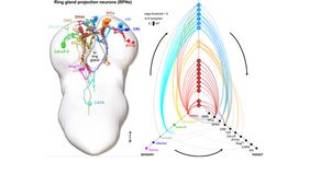
Neuroendocrine systems in animals maintain organismal homeostasis and regulate stress response. Although a great deal of work has been done on the neuropeptides and hormones that are released and act on target organs in the periphery, the synaptic inputs onto these neuroendocrine outputs in the brain are less well understood. Here, the Pankratz lab (first author: Dr. Sebastian Hückesfeld) has used the transmission electron microscopy reconstruction of a whole central nervous system in the Drosophila larva to elucidate the sensory pathways and the interneurons that provide synaptic input to the neurosecretory cells projecting to the endocrine organs. Predicted by network modeling, they also identify a new carbon dioxide-responsive network that acts on a specific set of neurosecretory cells and that includes those expressing corazonin (Crz) and diuretic hormone 44 (Dh44) neuropeptides. Their analysis reveals a neuronal network architecture for combinatorial action based on sensory and interneuronal pathways that converge onto distinct combinations of neuroendocrine outputs.
Publication:
Hückesfeld S, Schlegel P, Miroschnikow A, Schoofs A, Zinke I, Haubrich AN, Schneider-Mizell CM, Truman JW, Fetter RD, Cardona A, Pankratz MJ. Unveiling the sensory and interneuronal pathways of the neuroendocrine connectome in Drosophila. Elife. 2021 Jun 4;10:e65745. doi: 10.7554/eLife.65745.PMID: 34085637 Free PMC article.


